
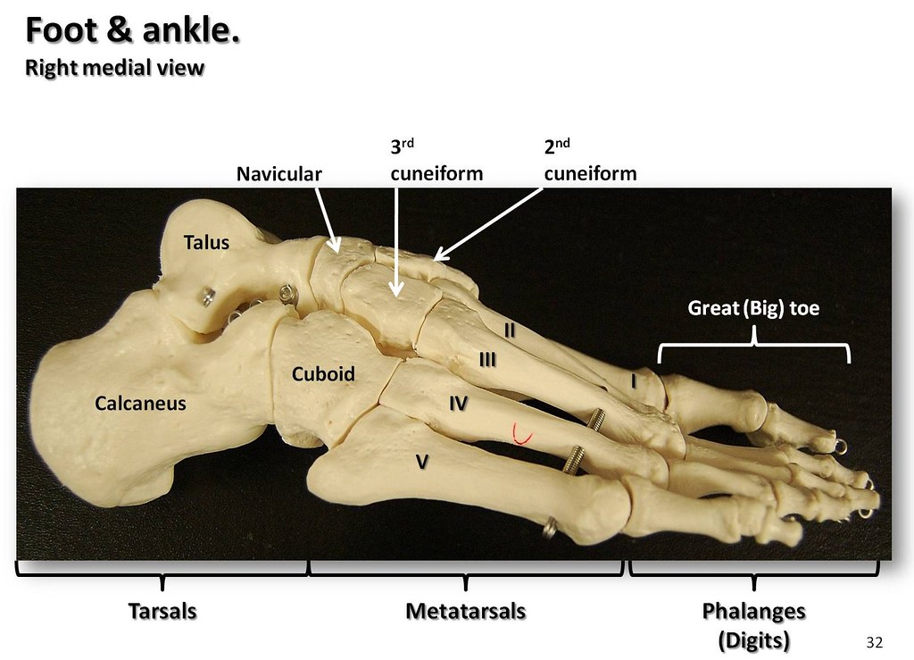
Bones of the ankle and foot. Bones of the ankle, Human body bones
Details. The original file was in Wavefront .OBJ format. The following is the original legend from the file: Foot Bones # # Courtesy of: # # Viewpoint Animation Engineering # 870 West Center # Orem, Utah 84057 # (801)224-2222 # 1-800-DATASET # $ Contributed to the FTP site at avalon.chinalake.navy.mil (129.131.31.11) # by Scott R. Nelson of Sun.

Ankle and Foot Pain Massage Therapy Connections
Foot bones labeling diagrams Foot bones labeled Foot bones unlabeled Sources + Show all Revise foot bone anatomy As you already know, there are 26 bones in the foot (FYI, that's almost a quarter of all the bones in the body!). But how are these distributed? The answer is between three distinct groups:
Diagrams of Foot 101 Diagrams
Foot and ankle anatomy consists of 33 bones, 26 joints and over a hundred muscles, ligaments and tendons. This complex network of structures fit and work together to bear weight, allow movement and provide a stable base for us to stand and move on. The foot needs to be strong and stable to support us, yet flexible to allow all sorts of complex.

Foot Anatomy, Gross Anatomy, Anatomy Art, Anatomy Drawing, Skeleton
Ankle anatomy The ankle joint, also known as the talocrural joint, allows dorsiflexion and plantar flexion of the foot. It is made up of three joints: upper ankle joint (tibiotarsal), talocalcaneonavicular, and subtalar joints. The last two together are called the lower ankle joint.
.jpg)
Foot Bone Diagram resource Imageshare
26 bones 33 joints more than 100 muscles, tendons, and ligaments Bones of the foot The bones in the foot make up nearly 25% of the total bones in the body, and they help the foot withstand.

Pin on Anatomny
1/2 Synonyms: Talocrural joint The foot is the region of the body distal to the leg that is involved in weight bearing and locomotion. It consists of 28 bones, which can be divided functionally into three groups, referred to as the tarsus, metatarsus and phalanges.
Bones of the foot and ankle, medial view with labels App… Flickr
Cuboid Medial cuneiform Intermediate cuneiform Lateral cuneiform Some people may be born with an extra navicular bone ( accessory navicular) beside the regular navicular bone, on the inside of the foot. This is a normal anatomical variation seen in around 2.5% of the entire population of the US. Metatarsal Bones

Foot & Ankle Bones
Skeletal System Bones of foot Bones of foot The 26 bones of the foot consist of eight distinct types, including the tarsals, metatarsals, phalanges, cuneiforms, talus, navicular, and cuboid.

Pin on Peopleanatomy
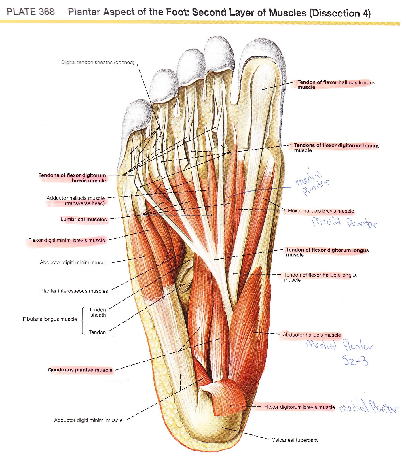
LABELED DIAGRAMS. Figure 1. Sections and Bones of the Foot A. Lateral (Left) B. Anterior (Right) Figure 2. Compartments of the Foot A. Cut Section through Mid-Foot. Figure 3. First Layer of the Foot A. Plantar View of Right Foot. Figure 4. Second Layer of the Foot A. Plantar View of Right Foot.

the muscles are labeled in this diagram
Common causes of foot pain include plantar fasciitis, bunions, flat feet, heel spurs, mallet toe, metatarsalgia, claw toe, and Morton's neuroma. If your feet hurt, there are effective ways to ease the pain. Some conditions specific to the foot can cause pain, less movement, or instability. Verywell / Alexandra Gordon.
Anatomy of human foot with labels on white background — ankle, leg
The 20-plus muscles in the foot help enable movement, while also giving the foot its shape. Like the fingers, the toes have flexor and extensor muscles that power their movement and play a large.

Ankle Bones Diagram koibana.info Foot anatomy, Anatomy bones, Ankle
Anatomy is a road map. Most structures in the foot are fairly superficial and can be easily palpated. Anatomical structures (tendons, bones, joints, etc) tend to hurt exactly where they are injured or inflamed.

anatomy of the foot Ballet News Straight from the stage bringing
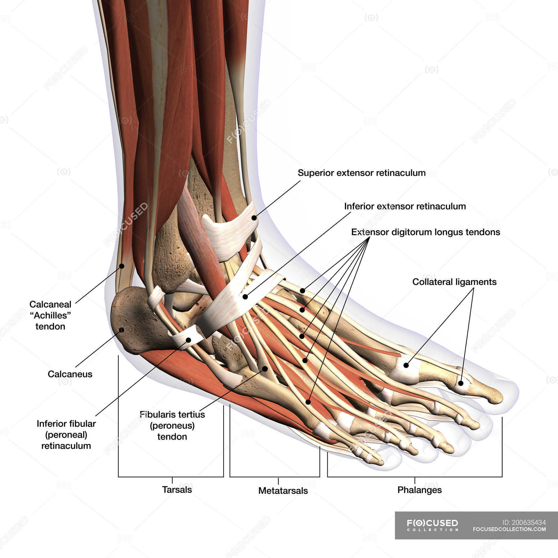
The extensor digitorum brevis is a small, thin muscle which lies underneath the long extensor tendons of the foot. Attachments: Originates from the calcaneus and inferior extensor retinaculum. It attaches onto the long extensor tendons of the medial four toes. Actions: Extension of the lateral four toes. Innervation: Deep fibular nerve.

How to have beautiful, healthy feet banish bunions and other
The diagram of bones in the ankle and foot is given below: Tarsal Bones The tarsal bones in the foot are located amongst tibia, metatarsal bones, and fibula. There are in all 7 bones, which fall under tarsal bones category. They are: Calcaneus or Calcaneum: To explain the term in layman's language, it is the heel bone in the skeletal system.

Anatomy Bones Learning Skeleton Feet Chapter 5 Appendicular Skeleton
The anatomy of the foot. The foot contains a lot of moving parts - 26 bones, 33 joints and over 100 ligaments. The foot is divided into three sections - the forefoot, the midfoot and the hindfoot. The forefoot. This consists of five long bones (metatarsal bones) and five shorter bones that form the base of the toes (phalanges).

Ankle Bones Diagram . Ankle Bones Diagram Ankle Diagrams Diagram Link
The Anatomy of Feet | Foot Diagrams, Physiology, and Pictures Made From The Molds Of Your Feet Active Designed for an active lifestyle. View Active Insoles Everyday Designed for normal day-to-day use. View Everyday Insoles Take Free Orthotics Quiz Table of Contents - Jump to section:
