
Labeled Diagram Of The Foot Teenage Lesbians
The muscles acting on the foot can be divided into two distinct groups; extrinsic and intrinsic muscles.. Extrinsic muscles arise from the anterior, posterior and lateral compartments of the leg. They are mainly responsible for actions such as eversion, inversion, plantarflexion and dorsiflexion of the foot. Intrinsic muscles are located within the foot and are responsible for the fine motor.

Parts of the feet and legs Grammar Tips
Foot Anatomy . There are many parts of the foot and all have important jobs. Each foot has 26 bones, over 30 joints, and more than 100 muscles, ligaments, and tendons. These structures work together to carry out two main functions:

Diagram of The Foot 101 Diagrams
In most two-footed and many four-footed animals, the foot consists of all structures below the ankle joint: heel, arch, digits, and contained bones such as tarsals, metatarsals, and phalanges; in mammals that walk on their toes and in hoofed mammals, it includes the terminal parts of one or more digits. The parts of a dog's hind foot and forefoot.
Notes on Anatomy and Physiology Using Imagery to Relax the Weight
The foot is one of the most complex parts of the body. It is made up of 26 bones connected by many joints, muscles, tendons, and ligaments. The foot is susceptible to many stresses. Foot problems can cause pain, inflammation, or injury. These problems can result in limited movement and mobility.
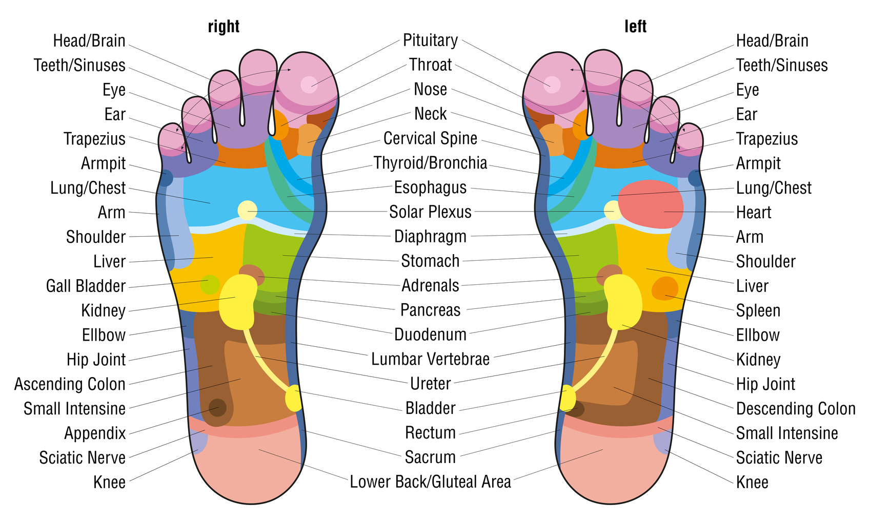
What is foot massage? Urban Blog
Parts of a Foot Your foot is made up of soft tissue and bones that work together to form a healthy, functioning, and pain-free foot. Muscles contract and relax to move the foot. Tendons are tough fibers that connect muscles to bones. Ligaments are fibrous strands that connect bones. Nerves travel throughout the foot, providing feeling.
Diagrams of Foot 101 Diagrams
The foot ( pl.: feet) is an anatomical structure found in many vertebrates. It is the terminal portion of a limb which bears weight and allows locomotion. In many animals with feet, the foot is a separate [clarification needed] organ at the terminal part of the leg made up of one or more segments or bones, generally including claws and/or nails.
Anatomy of human foot with labels on white background — ankle, leg
33 joints more than 100 muscles, tendons, and ligaments Bones of the foot The bones in the foot make up nearly 25% of the total bones in the body, and they help the foot withstand weight..
Bones of the human foot diagram 1142236 Vector Art at Vecteezy
The foot is an intricate part of the body, consisting of 26 bones, 33 joints, 107 ligaments, and 19 muscles. Scientists group the bones of the foot into the phalanges, tarsal bones, and.
Foot Pain Natural Relief and Herbal Remedies HubPages
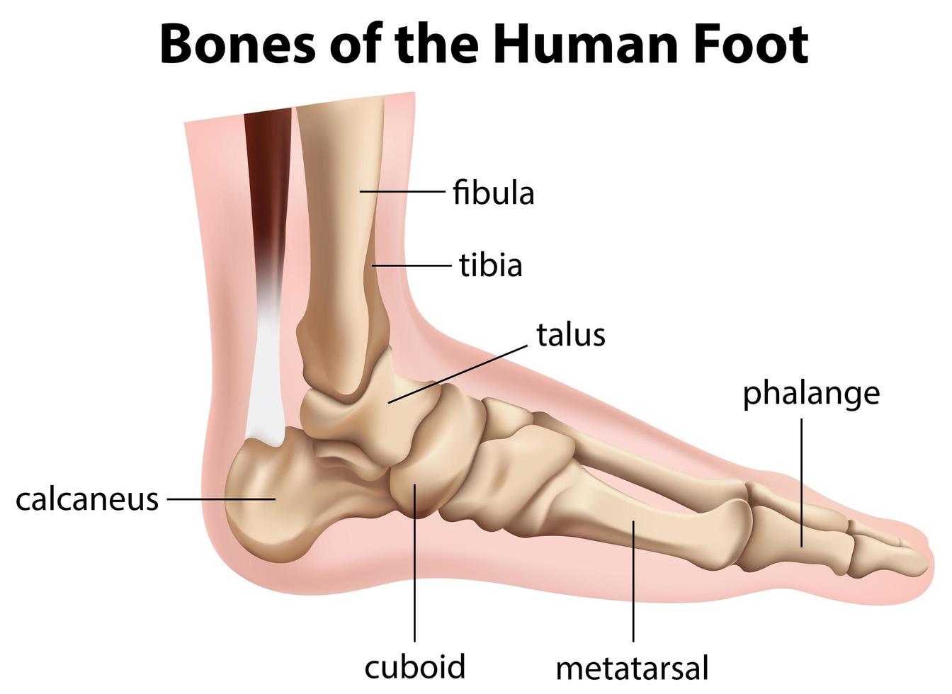
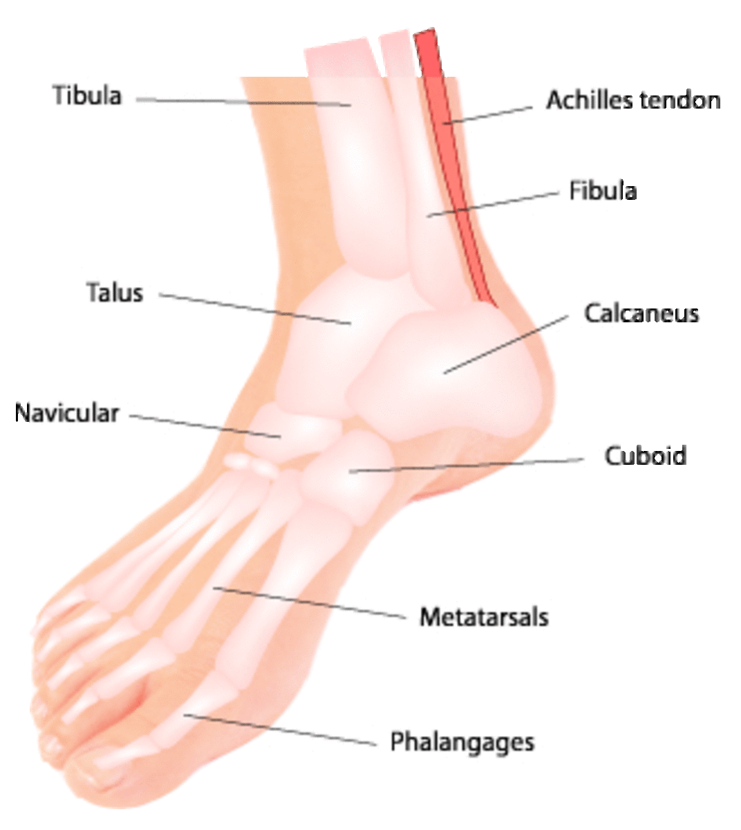
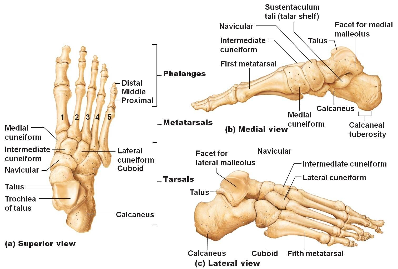
There are 26 bones in the foot, divided into three groups: Seven tarsal bones Five metatarsal bones Fourteen phalanges Tarsals make up a strong weight bearing platform. They are homologous to the carpals in the wrist and are divided into three groups: proximal, intermediate, and distal.
Lisfranc Injuries Core EM
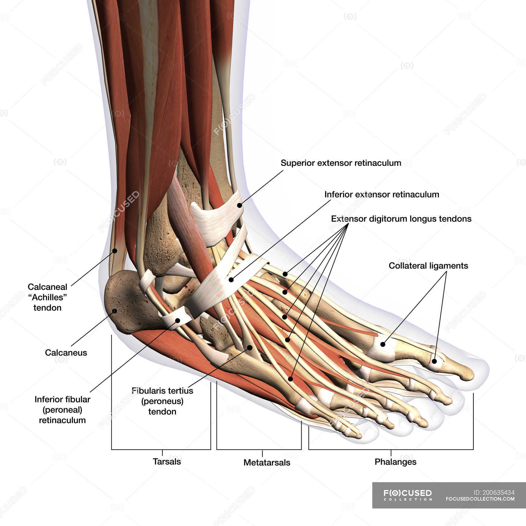
What are the Parts of the Foot Called? The feet are flexible structures of bones, joints, muscles, and soft tissues. They allow us to perform activities like walking, running, and jumping. Bones of the Foot The foot is divided into sections: The forefoot contains the five toes (phalanges) and the five longer bones (metatarsals).

Anatomy of the Foot and Ankle OrthoPaedia
These bones are arranged in two rows, proximal and distal. The bones in the proximal row form the hindfoot, while those in the distal row from the midfoot. Hindfoot. Talus. Calcaneus. The talus connects the foot to the rest of the leg and body through articulations with the tibia and fibula, the two long bones in the lower leg. Midfoot. Navicular.
Anatomical Charts and Posters Anatomy Charts Foot and Ankle
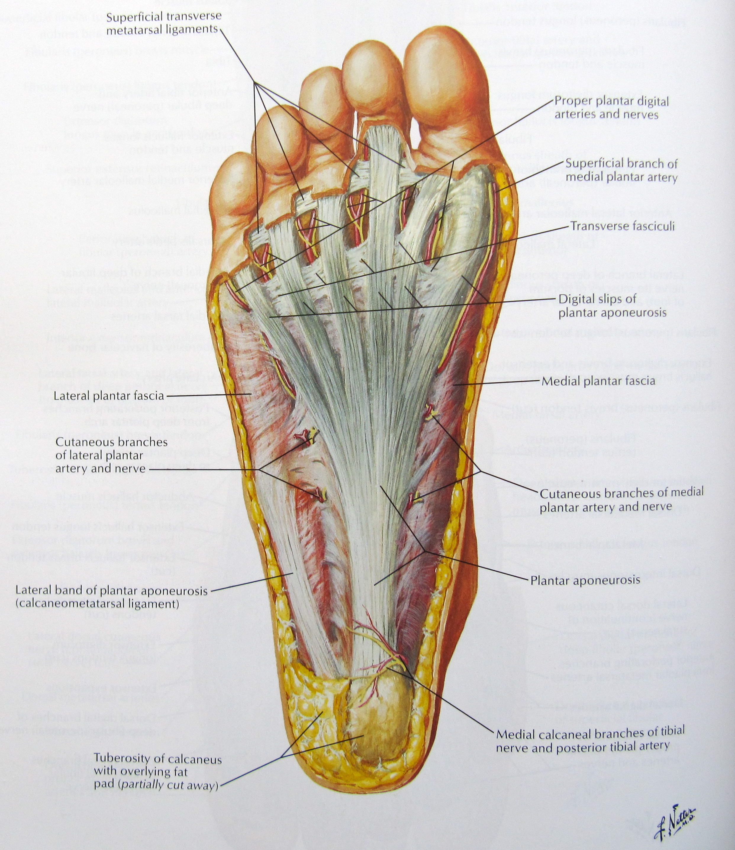
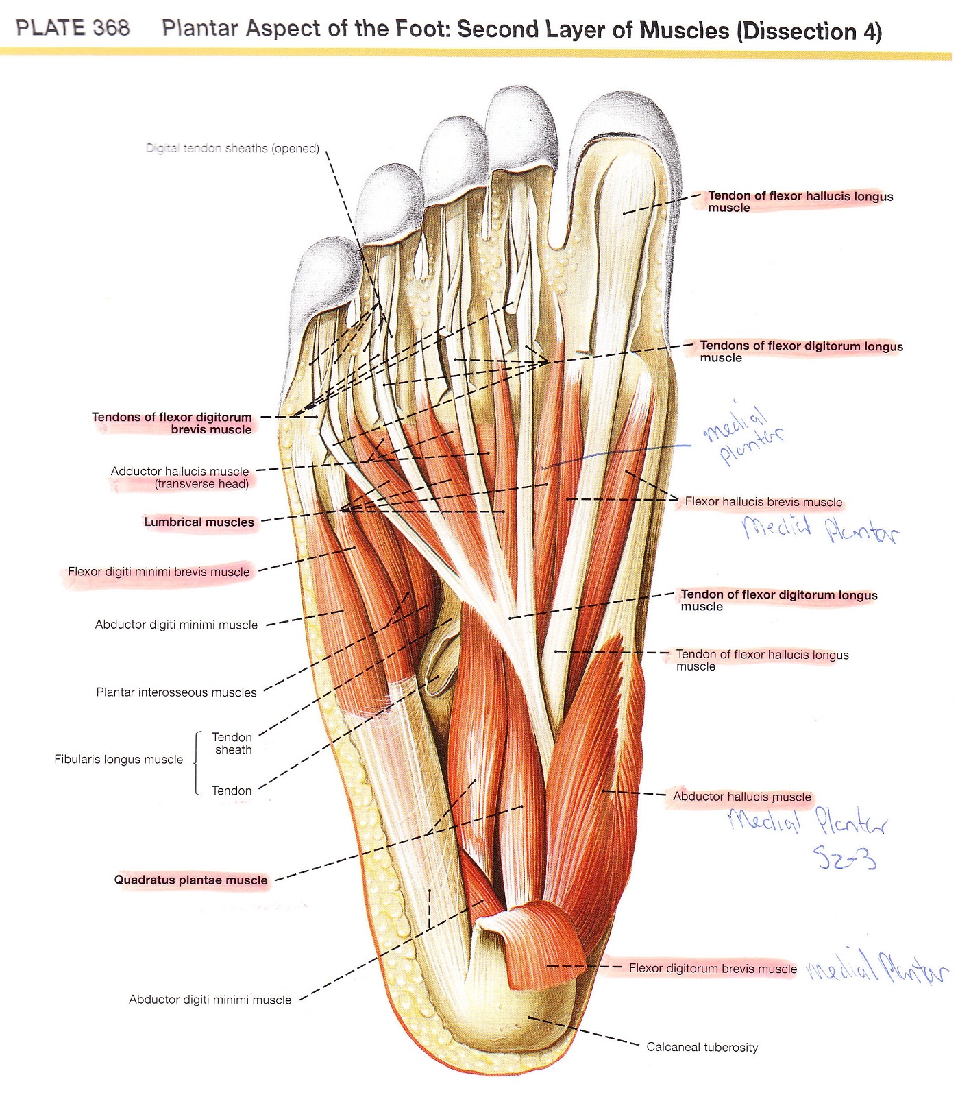
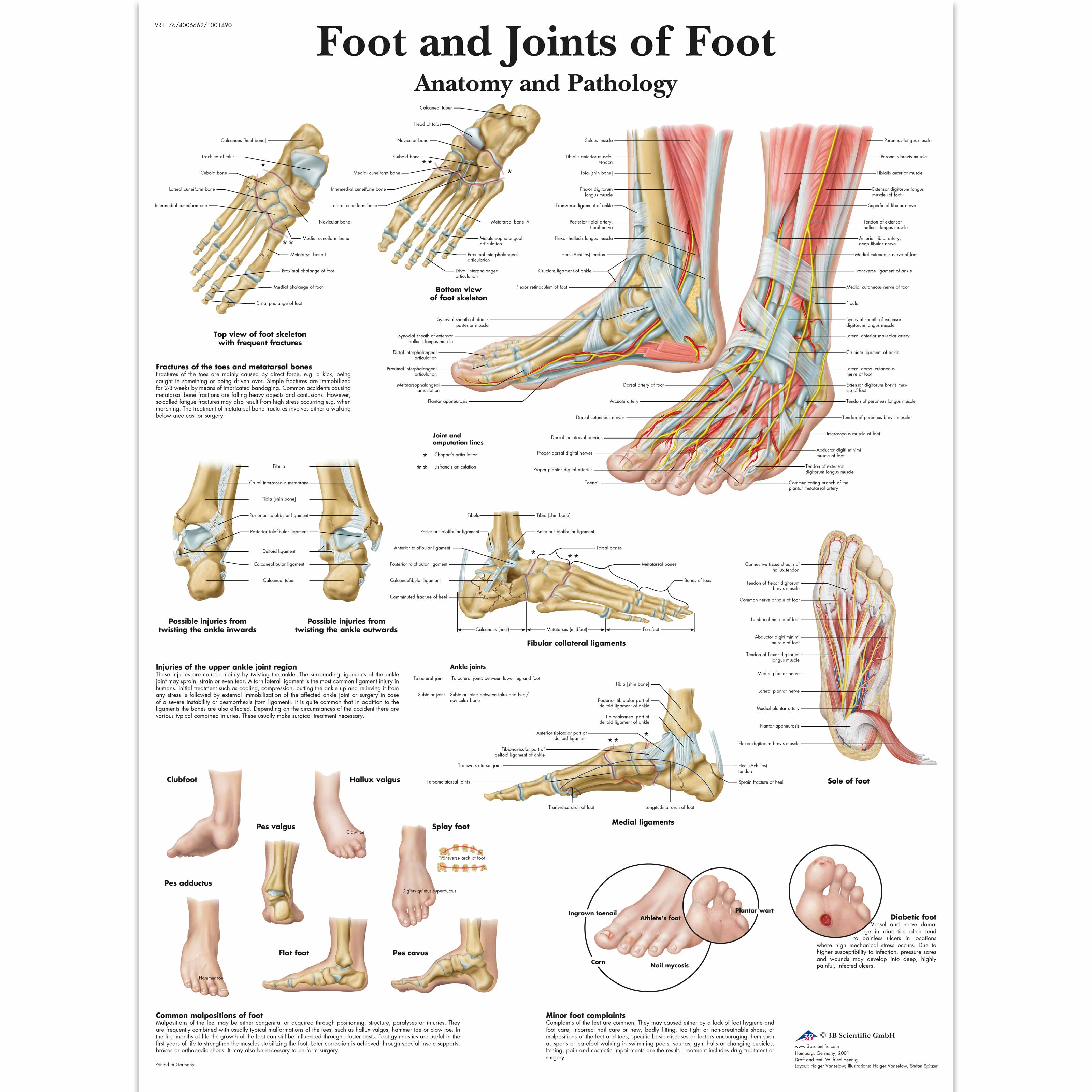
Anatomy of the Foot LABELED DIAGRAMS Figure 1. Sections and Bones of the Foot A. Lateral (Left) B. Anterior (Right) Figure 2. Compartments of the Foot A. Cut Section through Mid-Foot Figure 3. First Layer of the Foot A. Plantar View of Right Foot Figure 4. Second Layer of the Foot A. Plantar View of Right Foot Figure 5.

This chart shows foot and ankle bone and ligament anatomy, normal
Foot Anatomy The foot contains 26 bones, 33 joints, and over 100 tendons, muscles, and ligaments. This may sound like overkill for a flat structure that supports your weight, but you may not realize how much work your foot does!
Muscular Anatomy Of The Foot
The foot is one of the most complex parts of the body. It consists of 28 bones connected by many joints, muscles, tendons, and ligaments. The foot is prone to many types of injuries. Foot pain and problems can cause pain and inflammation, limiting movement. Muscles contract and relax to move the foot.

anatomy of the foot Ballet News Straight from the stage bringing
Synonyms: Talocrural joint The foot is the region of the body distal to the leg that is involved in weight bearing and locomotion. It consists of 28 bones, which can be divided functionally into three groups, referred to as the tarsus, metatarsus and phalanges.
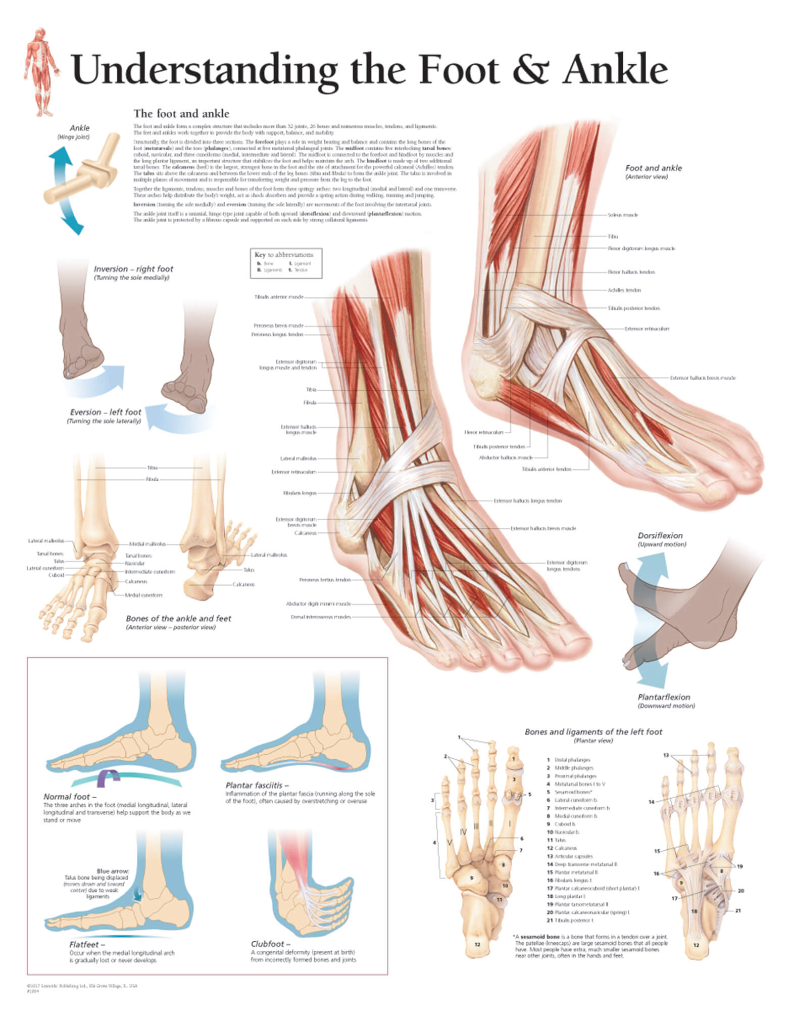
Understanding the Foot & Ankle Scientific Publishing
Bones of foot. The 26 bones of the foot consist of eight distinct types, including the tarsals, metatarsals, phalanges, cuneiforms, talus, navicular, and cuboid bones. The skeletal structure of.